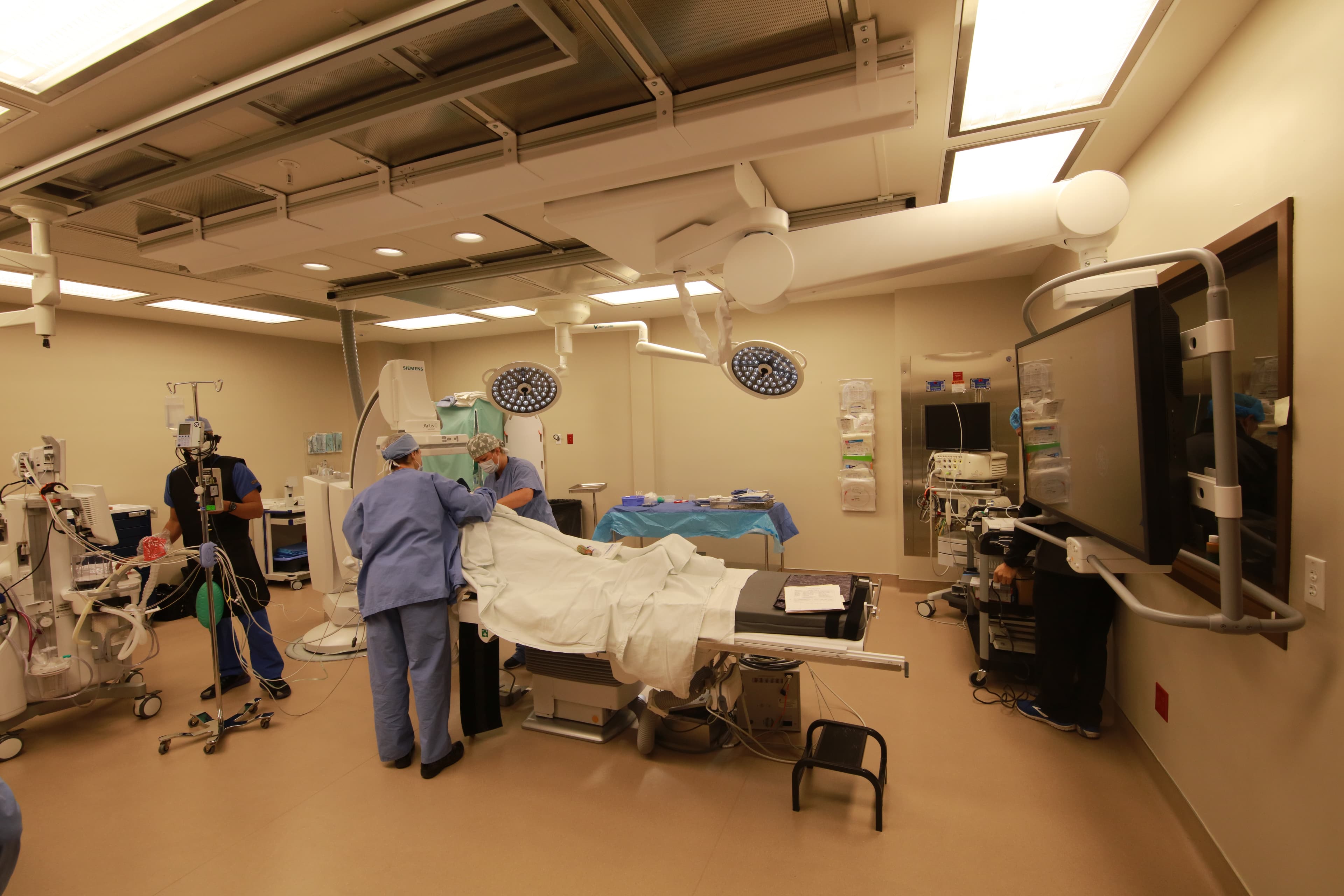
Coronary and peripheral catheter work, invasive haemodynamics, and imaging appropriate to an ambulatory surgery center.
Transesophageal Echocardiography (TEE)
High-resolution ultrasound of the heart taken from immediately behind it, used before and during rhythm procedures.
A slim ultrasound probe passed into the esophagus sits directly behind the heart, giving a far clearer view of the atria than an ultrasound taken through the chest wall. Before an ablation or a cardioversion it is used to confirm there is no clot in the left atrial appendage. It is performed under sedation and adds a short amount of time to the visit.
Commonly used for
- Pre-ablation clot exclusion
- Pre-cardioversion assessment
- Valve and atrial imaging
More about this procedure →Coronary Angiogram
X-ray imaging of the coronary arteries with contrast, to find out whether and where they are narrowed.
A catheter is passed to the origin of the coronary arteries, usually through the wrist, and contrast is injected while X-ray images are recorded. The result shows the arteries as a map: which are narrowed, by how much, and where. It is the test that decides between medication, stenting, and surgery, and where a narrowing is found that should be treated, treatment can often follow in the same visit.
Commonly used for
- Chest pain or shortness of breath on exertion
- Abnormal stress test
- Suspected coronary artery disease
More about this procedure →Coronary Intervention (Angioplasty and Stenting)
Opening a narrowed coronary artery with a balloon and holding it open with a stent.
A balloon is advanced over a fine wire to the narrowing and inflated to widen it, and a stent — a short metal mesh tube — is left behind to keep the artery open. Medicare added coronary angioplasty and stenting to the list of procedures covered in ambulatory surgery centers with effect from January 2020, for appropriately selected patients. Selection is the whole discipline here: this is elective, planned work in stable patients, not the treatment of a heart attack.
Commonly used for
- Stable angina
- Significant coronary narrowing on angiography
- Symptoms not controlled by medication
More about this procedure →Peripheral Angiogram
Contrast imaging of the arteries outside the heart, to establish where and how severely they are narrowed.
A catheter and contrast are used to image the arteries of the legs, pelvis, or elsewhere while X-ray images are recorded. It answers the questions a pulse examination and an ultrasound cannot: which segment is narrowed, over what length, and whether the vessel below it is open enough for treatment to help. Where a lesion is suitable, treatment can follow in the same visit.
Commonly used for
- Peripheral artery disease
- Claudication
- Suspected arterial narrowing in the leg
More about this procedure →Peripheral Intervention
Catheter treatment of narrowed arteries outside the heart, most often in the legs.
The same tools used in the coronary arteries — wires, balloons, and stents — are used to reopen narrowed arteries elsewhere in the body. In the legs, narrowing causes cramping pain on walking that eases with rest, and in more advanced disease it threatens wounds and healing. Imaging defines the disease first; treatment follows where it is appropriate and where the symptoms justify it.
Commonly used for
- Peripheral artery disease
- Claudication — leg pain on walking
- Non-healing wounds from poor arterial supply
More about this procedure →Right Heart Catheterization with Exercise
Direct measurement of the pressures inside the heart and lungs, at rest and during exercise.
A catheter passed through a vein measures pressure in the right atrium, right ventricle, pulmonary artery, and — through a balloon wedged in a small branch — the pressure on the left side of the heart. Measuring again during exercise matters because some patients have entirely normal pressures lying still and abnormal ones the moment they exert themselves, which is precisely when their symptoms occur. Resting measurements alone would call those patients normal.
Commonly used for
- Unexplained breathlessness on exertion
- Suspected pulmonary hypertension
- Heart failure with preserved ejection fraction
- Assessment before and after rhythm procedures
More about this procedure →