Skip to content
Outpatient coronary, structural, and rhythm procedures in Phoenix.Preparing for your procedure

Why HHC

A look inside

A center designed for one kind of work: short walking distances, imaging built into the room rather than a floor away, and a recovery area sized for the number of patients actually scheduled in a day.

A glimpse inside

Step inside Heart Health Center

A brief look through the doorway into our care area, with members of the team. This 34-second video has no audio.

Built for cardiac rhythm work

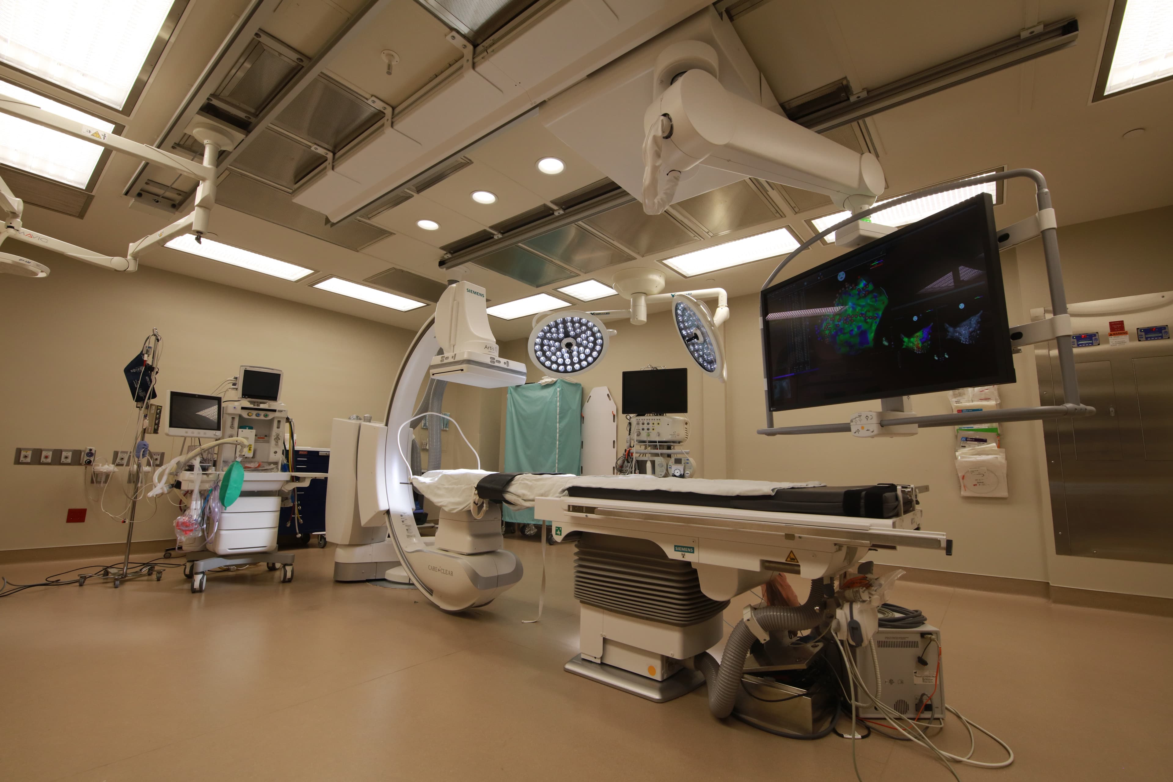
The procedure suites are equipped with fixed fluoroscopic imaging, full hemodynamic monitoring, and the three-dimensional mapping systems that electrophysiology requires — the same equipment class found in a hospital cardiac catheterization lab.

Around them, the layout is deliberately compact. Registration, pre-op, the procedure room, and recovery are steps apart, so a patient is never transported through unfamiliar corridors and a family member is never far from an update.

The center also maintains the equipment and staffing expected of complex electrophysiology care, including pericardiocentesis capability on site and defined emergency transfer arrangements with nearby hospitals.

Safety and quality in detail →

The center itself

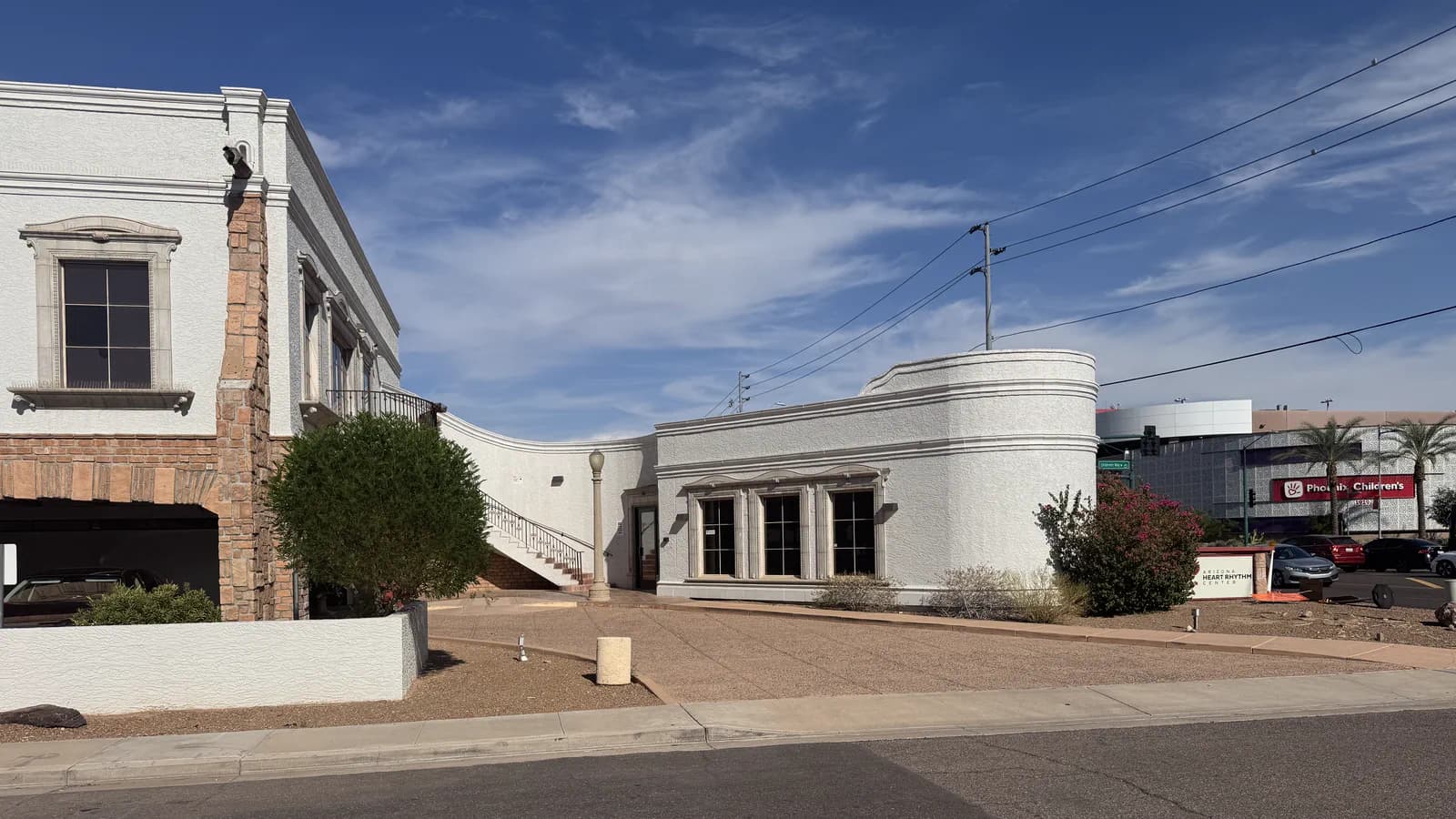
  • Heart Health Center seen from the street corner: a white two-story building with stone-faced pillars and arched windows, joined to a curved single-story rotunda wing. An exterior staircase sweeps up to a balcony entrance, and a covered drive passes beneath it.
    The center, from Thomas Road
  • The entrance elevation of the center. A covered drive runs under the building's first floor to a shaded doorway, so a patient can be dropped off out of the sun, with the exterior staircase and balcony above it.
    Covered entrance and drop-off
  • The curved single-story wing of the center, with tall arched windows and a stone cornice, set behind desert landscaping and flowering shrubs under a clear sky.
    The rotunda wing
  • A procedure suite with a fluoroscopic C-arm beside the procedure table, overhead surgical lights, and a large cardiac mapping display.
    Procedure suite
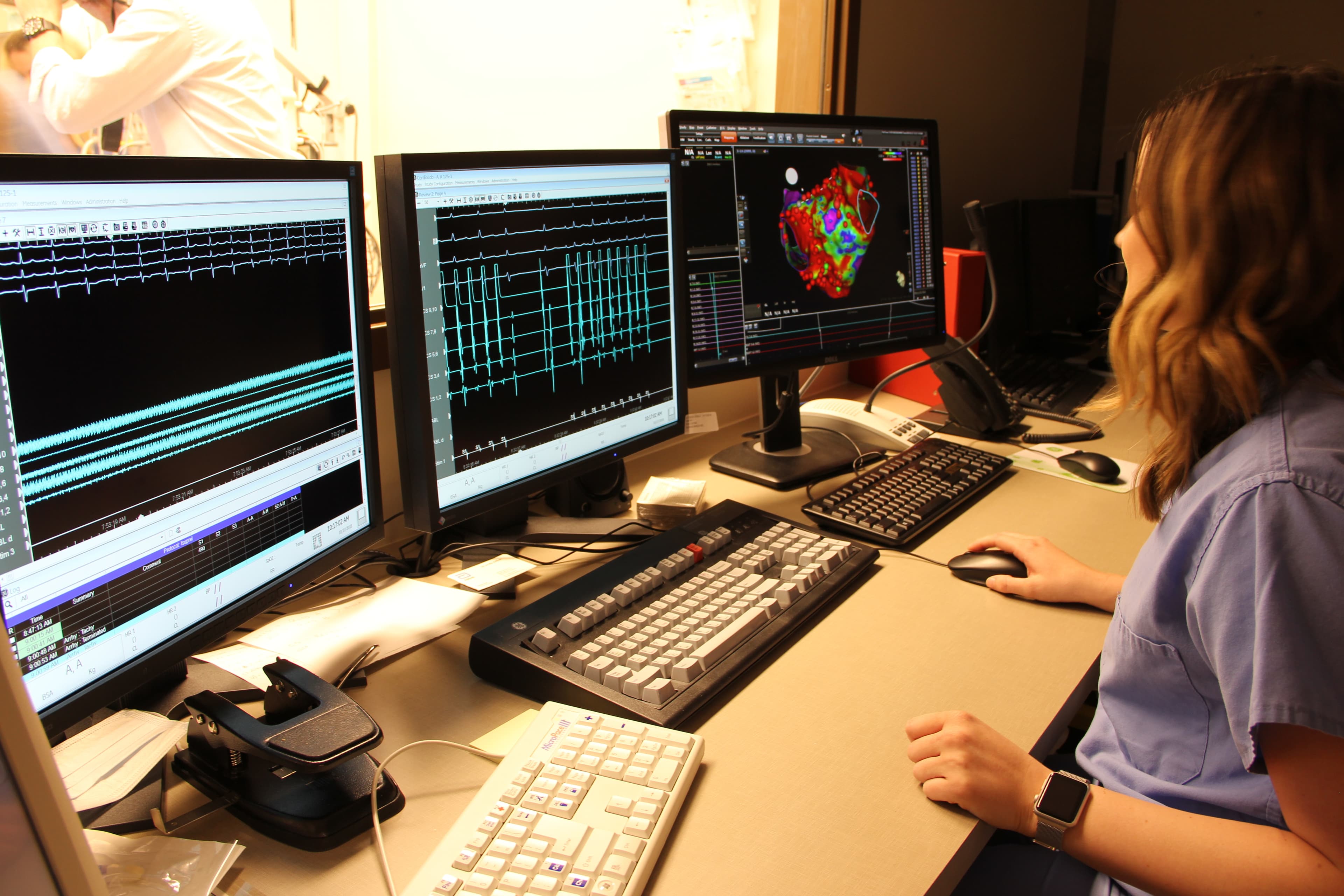
  • A team member at the monitoring workstation, viewing heart rhythm recordings and a three-dimensional cardiac map on multiple displays.
    Cardiac mapping and monitoring
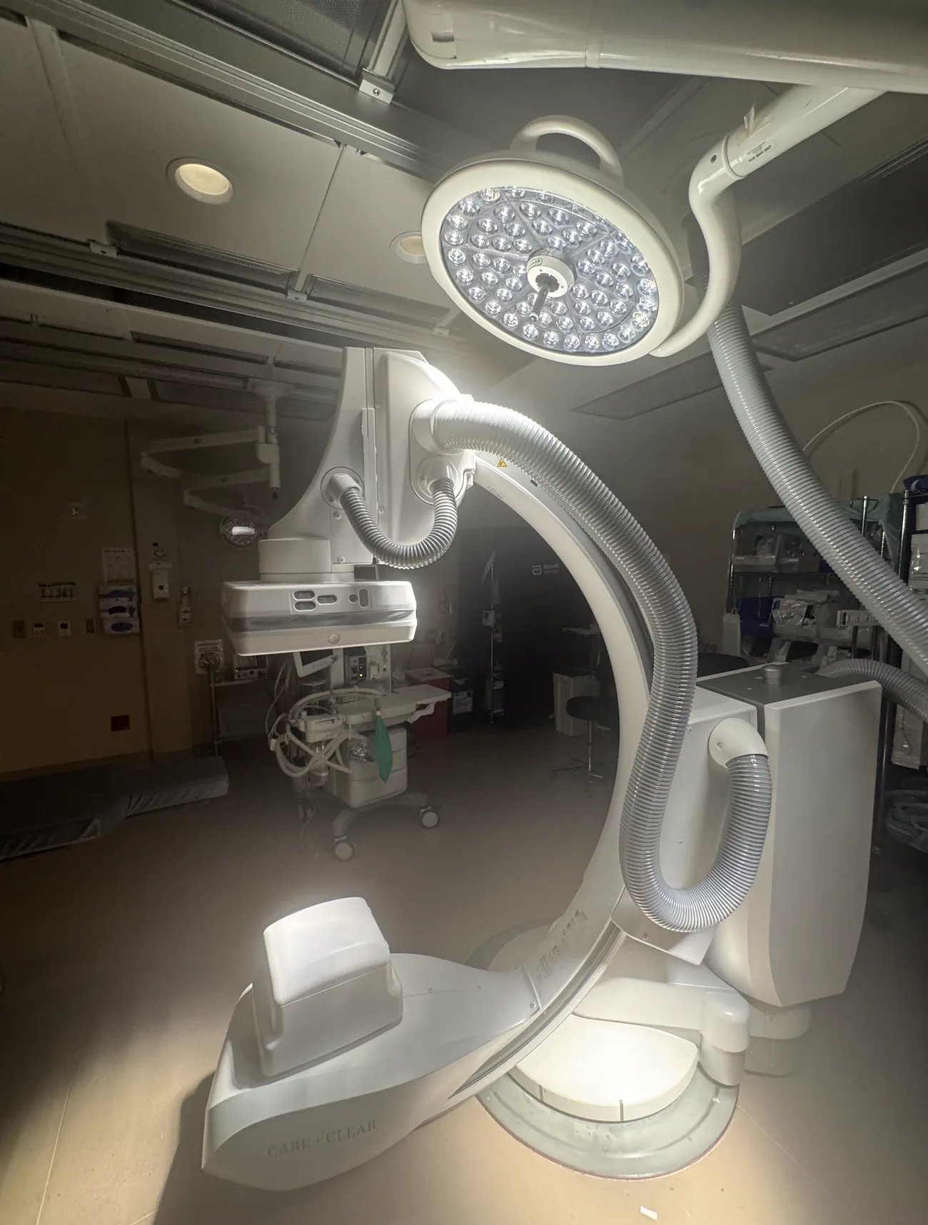
  • A close view of the fluoroscopic C-arm, its detector and X-ray source facing each other across the arc where the table sits, lit from above by a surgical light on an articulated boom.
    Fluoroscopic C-arm
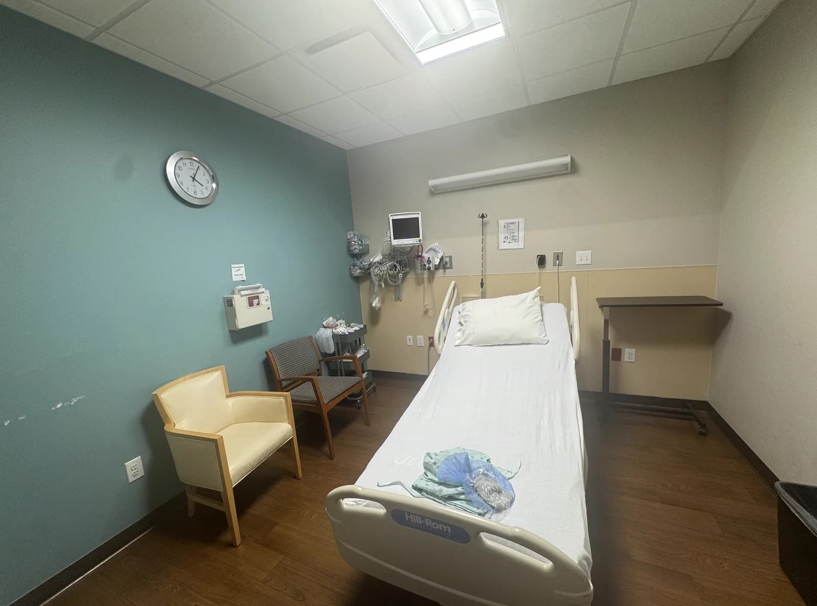
  • A private recovery bay: a hospital bed made up with a folded gown on it, a wall-mounted patient monitor and oxygen and suction outlets at the head of the bed, a clock on a teal accent wall, and a chair for a family member.
    Recovery bay
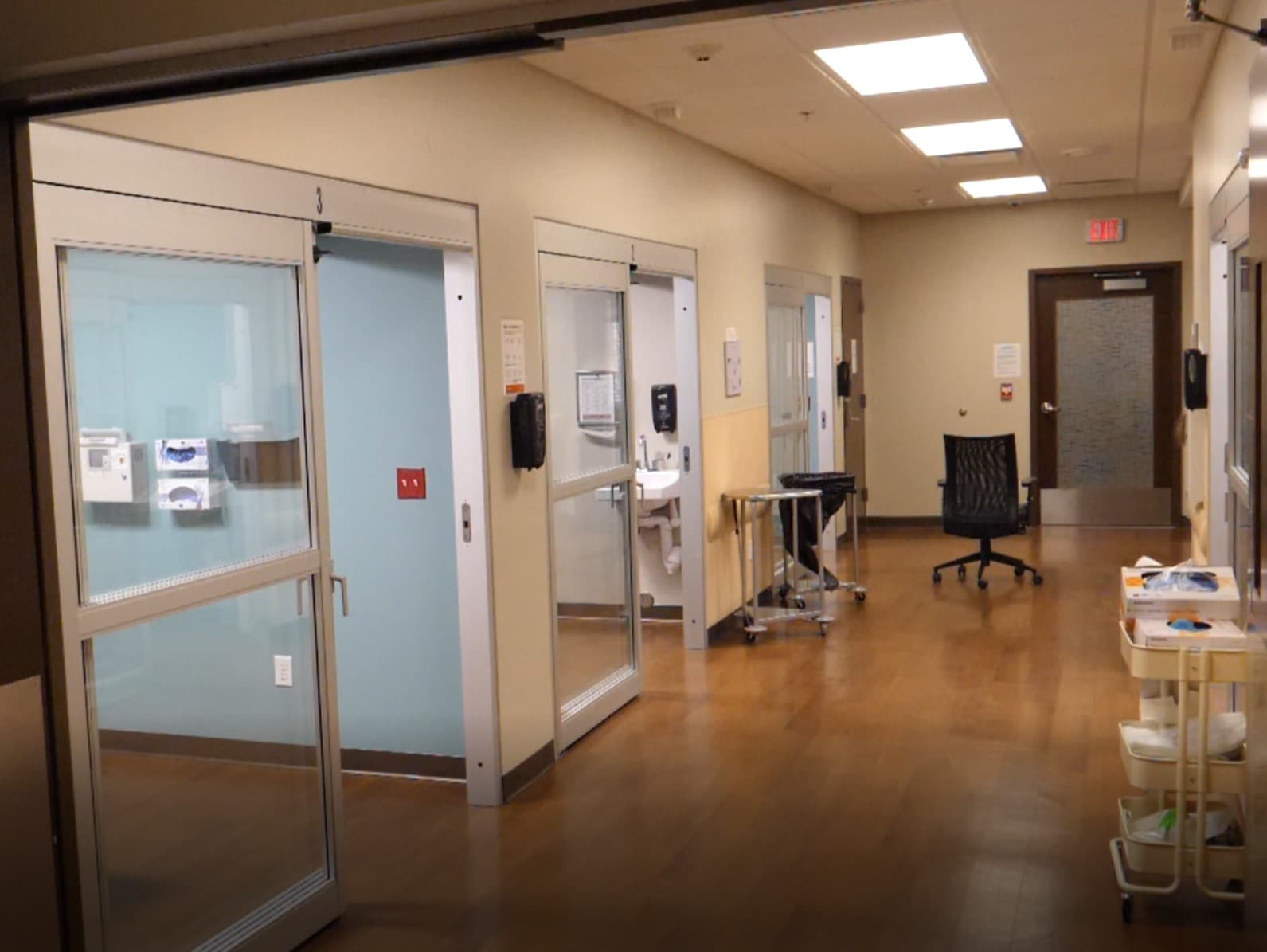
  • A hallway lined with recovery rooms, with sliding glass doors and privacy blinds.
    Recovery rooms
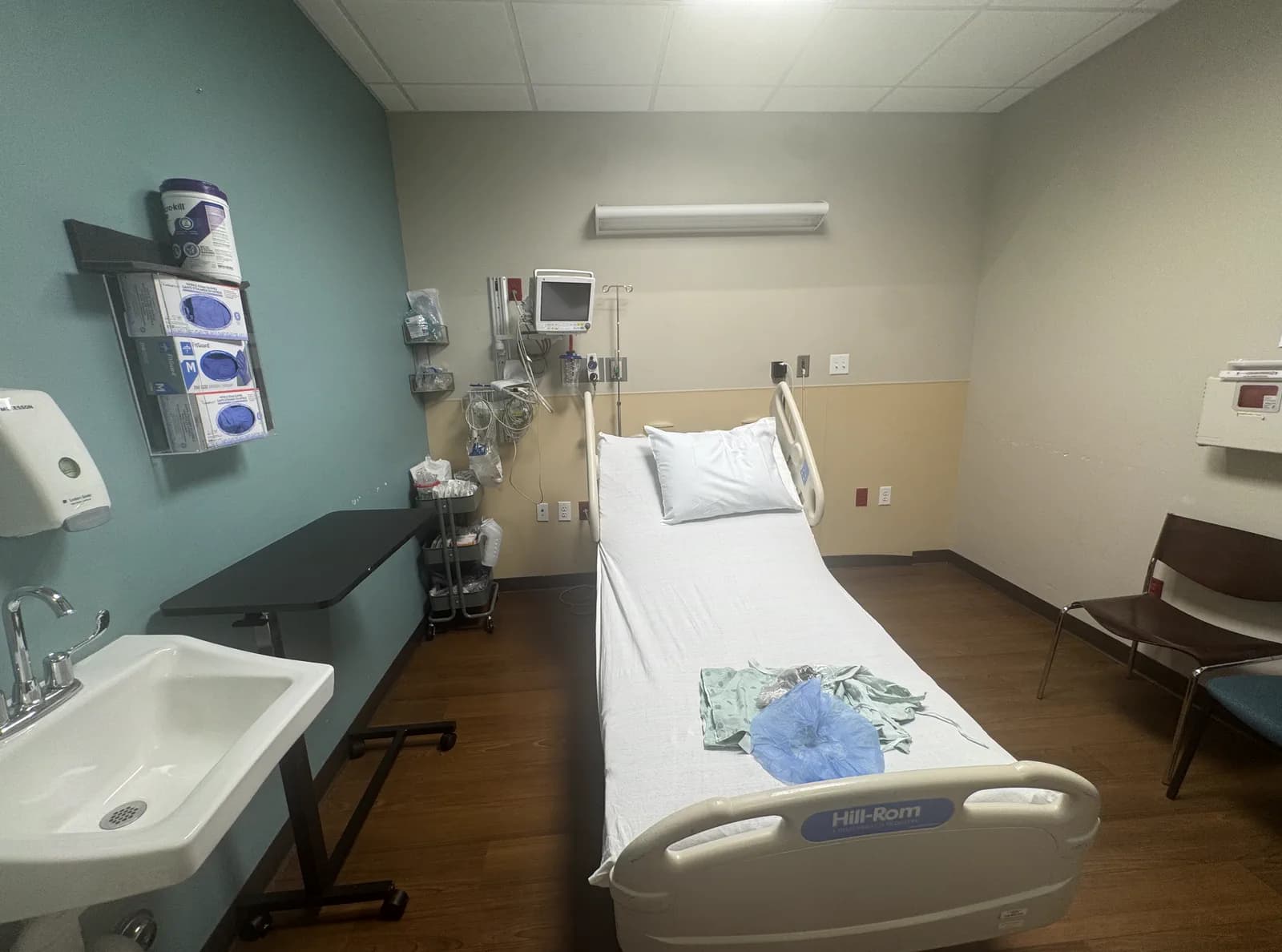
  • A pre-op bay with a bed, a wall-mounted monitor, a hand basin, a supply cart, and gloves and dressings stocked within reach at the bedside.
    Pre-op bay

Questions about your procedure?

Our team will confirm your date, walk you through preparation, and answer questions about insurance, sedation, and recovery.

Ask HHC