
Procedure
TEE and cardioversion
Two procedures, usually one visit. The echocardiogram is not a formality before the shock — it is what makes the shock safe to give that day.
The Two Halves
What each procedure does
One looks, one treats. They are scheduled together because the answer from the first decides whether the second happens.
The TEE — a clear look at the left atrium
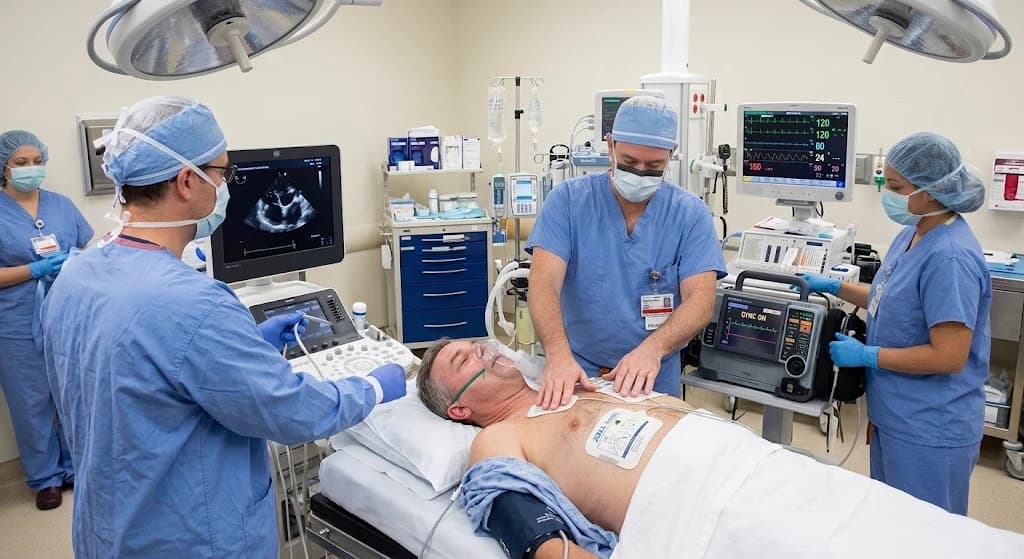
A transesophageal echocardiogram uses an ultrasound probe passed into the esophagus, which runs directly behind the heart. With no ribs or lung tissue in the way, it shows the left atrium and its appendage far more clearly than an ultrasound taken through the chest wall — clearly enough to say whether a clot is present.
The cardioversion — resetting the rhythm
Cardioversion delivers one brief electrical shock through pads on the chest, timed to the heart's own electrical cycle. It stops the disorganised activity for a fraction of a second, which is usually enough for the heart's natural pacemaker to take over again in a normal rhythm.
The Reason for the Order
Why the echocardiogram comes first
This is the part worth understanding before you consent, because it is the reason the visit is built the way it is.
- 01
Atrial fibrillation lets clots form
When the atrium fibrillates rather than contracts, blood moves sluggishly in the left atrial appendage — a small pouch off the left atrium. After roughly 48 hours of atrial fibrillation, or when nobody can say how long it has been going on, a clot may have formed there.
- 02
Restoring the rhythm can dislodge one
Cardioversion works by restoring organised contraction. If a clot is sitting in the appendage, that returning contraction is exactly what can push it out into the circulation, and from there to the brain. The shock itself is not the danger; the newly effective squeeze is.
- 03
So you either wait, or you look
The conventional route is at least three weeks of anticoagulation before the cardioversion, on the assumption that any clot has been treated. The alternative is to look directly with a TEE: if the appendage is clear, the cardioversion can go ahead in the same visit rather than three weeks later.
- 04
What looking actually finds
In the ACUTE trial, which randomised patients between the two strategies, a thrombus was found in 14% of those imaged — 76 of 619. Those patients were anticoagulated instead of shocked. That is the case for looking: roughly one patient in seven is not ready for a cardioversion that day, and imaging is what identifies them.
The Visit
What the day looks like
Both procedures happen under one sedation, in one room, in a single outpatient visit.
- 1
Before you come in
You will be told when to stop eating and drinking — the stomach needs to be empty for sedation. Your anticoagulation is reviewed in advance and is usually continued, not stopped. Arrange for a responsible adult to take you home; you will not be able to drive.
- 2
Sedation
Both procedures are done under sedation managed by the anesthesia team, so you will not be aware of either. The back of the throat is also numbed to make the probe comfortable to pass.
- 3
The TEE
The probe is passed into the esophagus and the left atrium and its appendage are examined. Your physician is looking specifically for thrombus, and also takes the opportunity to assess valve function and chamber size.
- 4
The decision point
If the appendage is clear, the cardioversion goes ahead straight away, while you are still sedated. If a clot is seen, the cardioversion is deliberately not performed — you are anticoagulated instead and the procedure is rescheduled. Being sent home without a shock is a good outcome on those days.
- 5
The cardioversion
Pads already placed on the chest deliver a single synchronised shock. Your rhythm is checked immediately afterwards, and if the first shock does not hold, a second at a higher energy may be given while you are still asleep.
- 6
Recovery and going home
You wake in the recovery bay and your rhythm is monitored for a defined period. A sore throat and a hoarse voice for a day or two are normal. Anticoagulation continues for at least four weeks afterwards regardless of what the TEE showed, because the atrium takes time to contract normally again. Most patients go home the same day.
Cardioversion restores a rhythm; it does not treat the reason the rhythm went wrong. When atrial fibrillation keeps coming back, that is usually the conversation that leads to ablation.
Risks
What can go wrong, and how likely it is
Serious complications are uncommon. These are the ones worth understanding before you consent, most serious first.
Injury to the esophagus
Passing the probe can scrape or, very rarely, tear the esophagus. Perforation is reported below 0.1% in large series, and overall morbidity from TEE is around 0.2% — but a perforation is serious when it happens, which is why the probe is passed under sedation by an operator who does this routinely.
Stroke
This is the risk the TEE exists to reduce, and imaging reduces it rather than removing it. A very small clot can escape detection, and the atrium's contraction does not return to normal immediately. That is why anticoagulation continues for at least four weeks after the cardioversion.
Rhythm problems just after the shock
The heart can beat slowly or pause briefly in the moments after cardioversion. You are monitored continuously through this window, and it is one of the reasons the recovery period is defined rather than left to judgement.
Sore throat, hoarseness, and difficulty swallowing
Common, expected, and short-lived — usually a day or two. Tell the team if swallowing is still uncomfortable after that, rather than waiting it out.
Skin irritation where the pads sit
Redness and occasionally a superficial burn on the chest where the pads were placed. It settles on its own.
The rhythm may not hold
Cardioversion restores a normal rhythm; it does not treat the reason the rhythm went wrong. Atrial fibrillation returns in a proportion of patients, sometimes within days. When it keeps returning, that is often the conversation that leads to ablation.
Sources
Questions about your procedure?
Our team will confirm your date, walk you through preparation, and answer questions about insurance, sedation, and recovery.