Skip to content
Outpatient coronary, structural, and rhythm procedures in Phoenix.Preparing for your procedure

Technology

Radiofrequency ablation

The precise tool. Where pulsed field ablation is built to isolate the pulmonary veins, radiofrequency places one small lesion exactly where the arrhythmia lives — which is what most rhythm problems outside atrial fibrillation actually need.

01

First ASC to perform SVT ablation with the Biosense CARTO Mapping System.

02

First ASC to perform AF ablation with the Biosense CARTO Mapping System.

03

First ASC to perform 1000 ablations.

The Procedure

How a radiofrequency ablation works

Four steps, none of which involve opening the chest. The whole visit is an outpatient one, and most patients go home the same day.

  1. Step 1

    A catheter, through a vein

    Access is through a vein in the groin. Nothing is opened surgically — the catheters are threaded up to the heart through the vessel, which is why this is a same-day procedure rather than an operation.

  2. Step 2

    The circuit is mapped first

    Before any energy is delivered, mapping systems such as CARTO and Abbott EnSite build a three-dimensional map of the chamber and track where the catheter tip sits inside it. For many arrhythmias the map is the hard part: find where the abnormal circuit runs, and the treatment follows.

  3. Step 3

    Radiofrequency energy heats a small volume of tissue

    The catheter tip delivers radiofrequency current, which warms a few millimetres of heart muscle until the cells can no longer carry an electrical signal. The tip is irrigated with saline so the surface stays cool and the heating happens in the tissue rather than at the electrode.

  4. Step 4

    The lesion is checked, not assumed

    The catheter reports how firmly it is pressed against the tissue, and the delivered dose is recorded on the map lesion by lesion. The operator can see which points are underdosed rather than inferring it from the outcome — which is what makes a line durable.

Conditions

What radiofrequency ablation treats

These are the rhythms where a discrete, well-placed lesion is the treatment — and where a catheter designed around the pulmonary veins would be the wrong instrument.

AV nodal reentrant tachycardia (AVNRT)
The most common regular fast rhythm in otherwise healthy hearts, caused by a small reentry circuit beside the AV node. It is the textbook radiofrequency target — a discrete, well-localised problem.
Accessory pathway tachycardia (AVRT, WPW)
An extra electrical connection between atrium and ventricle. Ablation is aimed at the pathway itself, which has to be located precisely before it can be treated.
Typical atrial flutter
A large reentry circuit in the right atrium that depends on a defined strip of tissue, the cavotricuspid isthmus. A line of radiofrequency lesions across that isthmus interrupts the circuit.
Atrial tachycardia
A focal source elsewhere in the atrium. Mapping identifies where the earliest activation begins; the ablation is aimed there.
Selected atrial fibrillation cases
Most AF ablation here is done with pulsed field. Radiofrequency is used where the anatomy, prior ablation, or an additional target makes a titratable focal lesion the better tool.

≈98%

Long-term freedom from arrhythmia after AVNRT ablation

≈92%

Long-term freedom from arrhythmia after accessory pathway ablation

0.5%

Overall complication rate across more than 10,000 SVT ablations, 2005–2020

These figures come from published series, not from this center. They describe what the procedure achieves in general, and they are not a prediction about any individual patient.

Choosing the Energy

Why radiofrequency and not pulsed field

This center adopted pulsed field ablation early and uses it for most atrial fibrillation work. That is a reason to be specific about when radiofrequency is chosen instead, rather than vague.

Arrhythmias that are not the pulmonary veins

Supraventricular tachycardia, atrial tachycardia, typical atrial flutter, and accessory pathways are focal or narrowly anatomical problems. They need a discrete, precisely placed lesion — not a circumferential one designed for a vein. Radiofrequency delivered through a steerable focal catheter is the direct tool for that, and pulsed field catheters are not shaped for it.

When the circuit has to be found first

Scar-related and atypical arrhythmias are a mapping problem before they are an ablation problem. CARTO 3 builds activation and voltage maps that show where the circuit actually runs, so the ablation is aimed at a target the operator has identified rather than at an anatomical assumption.

When lesion dose needs to be verifiable

Contact force and per-lesion dose tracking turn 'the line looks complete' into something measurable. Where a durable focal lesion in one specific place is the whole objective, that feedback is worth more than speed.

Which energy a given case gets is decided by the operator, on the anatomy and the arrhythmia in front of them — not by which machine the center happens to own. For atrial fibrillation specifically, see pulsed field ablation and AFib ablation.

Platform

The system in use here

Mapping and ablation on one platform, from the vendor whose mapping system is the most widely used in electrophysiology.

Biosense Webster (Johnson & Johnson MedTech)

CARTO 3 System

THERMOCOOL SMARTTOUCH SF catheter

Three-dimensional electroanatomical mapping paired with contact-force radiofrequency ablation — the platform of choice when the target is a discrete focus rather than a vein.

CARTO 3 builds a three-dimensional map of the chamber and tracks the catheter tip within it, so the operator localises the circuit before delivering any energy. The THERMOCOOL SMARTTOUCH SF catheter is a 7.5 F irrigated radiofrequency catheter that reports the actual contact force between tip and tissue in real time — the variable that most determines whether a lesion is durable. Its porous tip cools uniformly at half the irrigation flow of earlier designs, which matters over a long case. Lesion-by-lesion dose is tracked on the map, so the operator can see which parts of a line are underdosed rather than inferring it.

What it changes

  • Maps the arrhythmia circuit before any energy is delivered
  • Reports real-time tip-to-tissue contact force, the main determinant of lesion durability
  • Records delivered dose per lesion on the map, exposing gaps in a line
  • Titratable focal lesions — the right tool away from the pulmonary veins

FDA status

FDA approved for drug-refractory paroxysmal atrial fibrillation; Biosense Webster announced the US launch of the THERMOCOOL SMARTTOUCH SF catheter in August 2016.

Pivotal trial

SMART-AF and SMART-SF — the trials establishing that contact-force feedback keeps the operator inside a target force range, with procedure times and outcomes that did not come at the cost of safety.
Mapping

What the map actually shows

Three views of the same left atrium, from a case performed at this center. Each answers a different question, and together they are how the circuit is found before any energy is delivered.

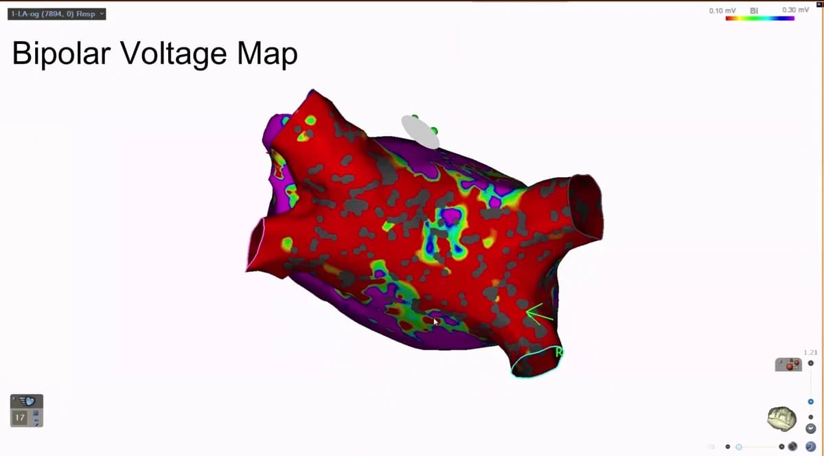
A bipolar voltage map of the left atrium on the CARTO 3 system. Most of the surface is red, marking low-amplitude tissue, with purple areas of preserved signal and scattered grey gaps where no point was taken.

Bipolar voltage

Signal strength across the chamber wall. Purple is healthy muscle carrying a normal signal; red marks tissue whose signal has dropped away. Before anything is treated, this is how the operator sees which parts of the atrium are still electrically alive.

CARTO 3 · Biosense Webster. From a case performed at Heart Health Center.

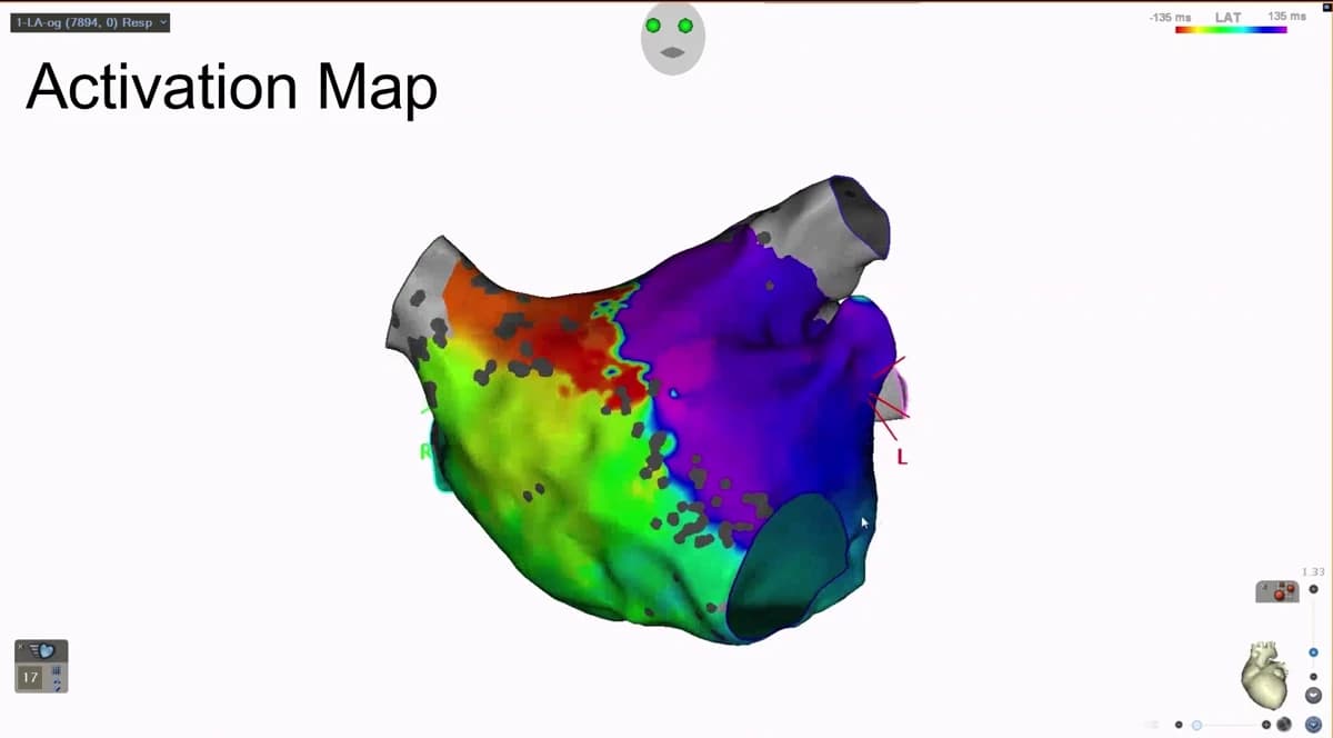
An activation map of the left atrium on the CARTO 3 system, coloured from red through the spectrum to purple to show the order in which regions of the chamber are electrically activated.

Activation timing

The same chamber, coloured by time instead of voltage. Red activates earliest and purple latest, so the colour sequence traces the path the electrical wavefront actually takes — which is what identifies the circuit driving the arrhythmia.

CARTO 3 · Biosense Webster. From a case performed at Heart Health Center.

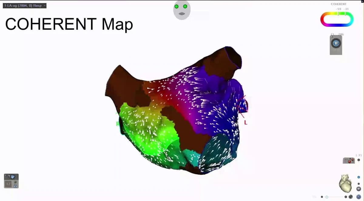
A CARTO 3 COHERENT map of the left atrium with white arrows across the surface indicating the direction and speed of electrical conduction.

Conduction direction

Every white arrow is the direction conduction is travelling at that point. Where the arrows converge, collide, or slow, the circuit has a vulnerable point — and that is where the ablation is aimed.

CARTO 3 · Biosense Webster. From a case performed at Heart Health Center.

Risks

What can go wrong, and how likely it is

Radiofrequency ablation is a safe procedure in experienced hands, and it is still a procedure. These are the complications worth understanding before you consent to one.

Injury to the conduction system

Ablating close to the AV node — which is where the target sits in AVNRT and in septal accessory pathways — carries a risk of heart block that would require a pacemaker. Published series put this at up to about 1%. It is the reason these ablations are done slowly, with the catheter position confirmed before energy is delivered.

Bleeding and vascular injury at the access site

The groin puncture can bruise, bleed, or rarely damage the vessel. Vascular complications occurred in about 0.1% of a large SVT ablation series.

Fluid around the heart

A catheter can perforate the wall of the heart and allow blood to collect in the sac around it. Cardiac tamponade was seen in about 0.3% of that same series, and it is managed by draining the fluid.

The risks specific to heating tissue

Because radiofrequency works by temperature, it can affect structures next to the heart — the esophagus behind the left atrium, the phrenic nerve beside the right pulmonary veins, and the veins themselves. These complications are rare, and avoiding them is the reason left atrial work here is usually done with pulsed field instead.

Stroke

As with any catheter procedure inside the heart, a clot can form and travel. Anticoagulation during and after the procedure is managed to reduce that risk.

Questions about your procedure?

Our team will confirm your date, walk you through preparation, and answer questions about insurance, sedation, and recovery.

Ask HHC