
Procedure
Electrophysiology study
The diagnostic step. Before an arrhythmia can be treated precisely it has to be identified precisely — which means recording the heart’s electrical signals from the inside, and bringing the rhythm on deliberately to see how it works.
The Study
What an EP study actually does
Two things a monitor cannot: it listens from inside the chambers, and it can start the rhythm on purpose.
It records from inside the heart
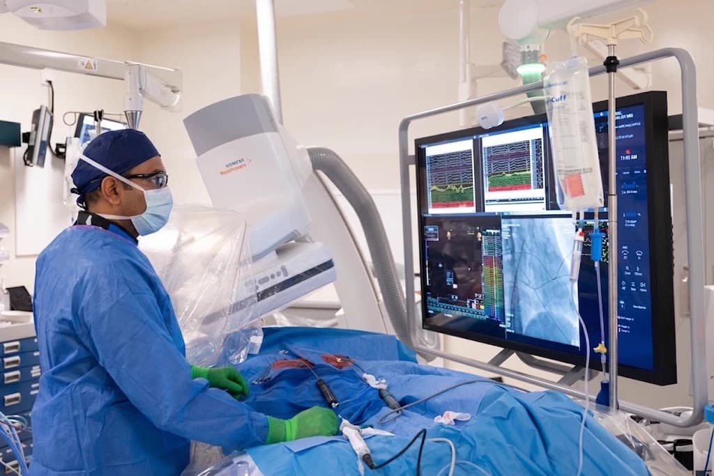
A surface ECG reads the heart's electrical activity through the chest wall, muscle, and lung. An electrophysiology study places sensing electrodes directly against the inside of the chambers, on catheters advanced through a vein at the groin. The signals it records are orders of magnitude more detailed, and they are local — they say which part of the heart did what, and in what order.
It can bring the arrhythmia on deliberately
An arrhythmia that comes and goes is difficult to diagnose because it is rarely present when anyone is recording. During a study the heart is paced in defined sequences to reproduce the rhythm under controlled conditions, with the team watching and ready. Provoking it on purpose is what allows its mechanism to be identified rather than guessed at.
Indications
Why a study gets ordered
A study is not a screening test. It is done when a specific question needs an answer that non-invasive testing has not given.
- Palpitations without an answer
- When an ECG and a monitor have not caught the rhythm but the symptoms are real and recurring, a study can reproduce it and name it.
- A known arrhythmia that needs locating
- Where the diagnosis is already established, the study defines exactly which tissue is responsible — which is the information an ablation needs to be precise.
- Unexplained fainting
- When syncope remains unexplained after non-invasive testing, particularly in someone with a previous heart attack or scarred heart muscle, a study can establish whether an arrhythmia is the cause.
- Assessing the conduction system
- Where a slow heart rate or heart block is suspected, the study measures how well each part of the conduction pathway is working, and how much reserve is left.
The Visit
What the day looks like
One sedation, one visit. Where an arrhythmia is found and can be treated, the treatment usually happens in the same session.
- 1
Before you come in
You will be told when to stop eating and drinking. Antiarrhythmic medication is often held for a period beforehand — deliberately, because a drug that suppresses the rhythm also prevents the study from finding it. Do not stop anything on your own; you will be given specific instructions. Arrange for someone to drive you home.
- 2
Sedation and access
The study is performed under sedation managed by the anesthesia team. A vein in the groin is accessed and the catheters are advanced to the heart. Nothing is opened surgically, and the access itself is a needle puncture.
- 3
Baseline recording
Before anything is provoked, the electrical intervals are measured at rest — how long the signal takes to travel from the atrium through the conduction system to the ventricle. Abnormalities here can be the diagnosis on their own.
- 4
Provoking the rhythm
The heart is then paced in defined sequences to try to start the arrhythmia. If it starts, its pattern on the recordings identifies the mechanism and the circuit. Occasionally a medication is given to make an elusive rhythm easier to provoke.
- 5
Treating it in the same visit
If an arrhythmia is induced and it is one that can be ablated, treatment usually follows immediately while the catheters are already in place. This is discussed and consented in advance, so the decision is not being made for the first time while you are asleep.
- 6
Recovery and going home
The catheters come out, the access site is closed, and you rest flat while it seals and your rhythm is monitored. Most patients go home the same day with written instructions and a follow-up plan.
Results
What the possible answers mean
Three outcomes, and the middle one is the one worth reading carefully.
The arrhythmia is induced and identified
The most useful result. The mechanism is known, the responsible tissue is localised, and in most cases it can be treated during the same visit.
Nothing is induced
Reassuring, and worth understanding precisely: a normal study means no arrhythmia could be provoked that day. It does not prove that none exists. A negative study cannot rule out an arrhythmic cause of fainting, so if your symptoms continue, they still need reporting rather than dismissing.
The conduction system is the problem
Sometimes the finding is not a fast rhythm but a failing pathway — the signal taking too long, or intermittently not arriving. That points towards a pacemaker rather than an ablation.
Where the study finds a circuit that can be treated, that treatment is usually radiofrequency ablation in the same session. For the broader picture of what an electrophysiologist does, see cardiac electrophysiology.
Risks
What can go wrong, and how likely it is
Serious complications are uncommon. These are the ones worth understanding before you consent, most serious first.
Fluid around the heart
A catheter can perforate a chamber wall and allow blood to collect in the sac around the heart. This is uncommon, and it is the complication the team is most alert to; it is managed by draining the fluid.
The arrhythmia that is provoked on purpose
Starting the rhythm is the point of the study, and most provoked rhythms are stopped by pacing. Occasionally one does not settle and a synchronised shock is used to restore a normal rhythm while you are still sedated. The defibrillator is in the room and connected before the study begins, for exactly this reason.
Injury to the conduction system
Catheters sit close to the heart's own wiring, and ablating near the AV node carries a risk of heart block that would require a pacemaker. Where the target is in that region, this is discussed specifically before you consent.
Bleeding and vascular injury at the access site
Bruising is common and settles. Less often the vessel is damaged or a collection of blood forms and needs attention.
Stroke
Any catheter inside the heart can allow a clot to form and travel. Anticoagulation during the procedure is managed to reduce that risk.
Radiation from X-ray imaging
Fluoroscopy is used to see the catheters. Three-dimensional mapping reduces how much of it is needed, and exposure is kept to the minimum that lets the study be done safely.
Sources
Questions about your procedure?
Our team will confirm your date, walk you through preparation, and answer questions about insurance, sedation, and recovery.